50+ Neck Anatomy Thyroid Cartilage. Normal ultrasound anatomy and technique. In the midline, alongside the base of the.
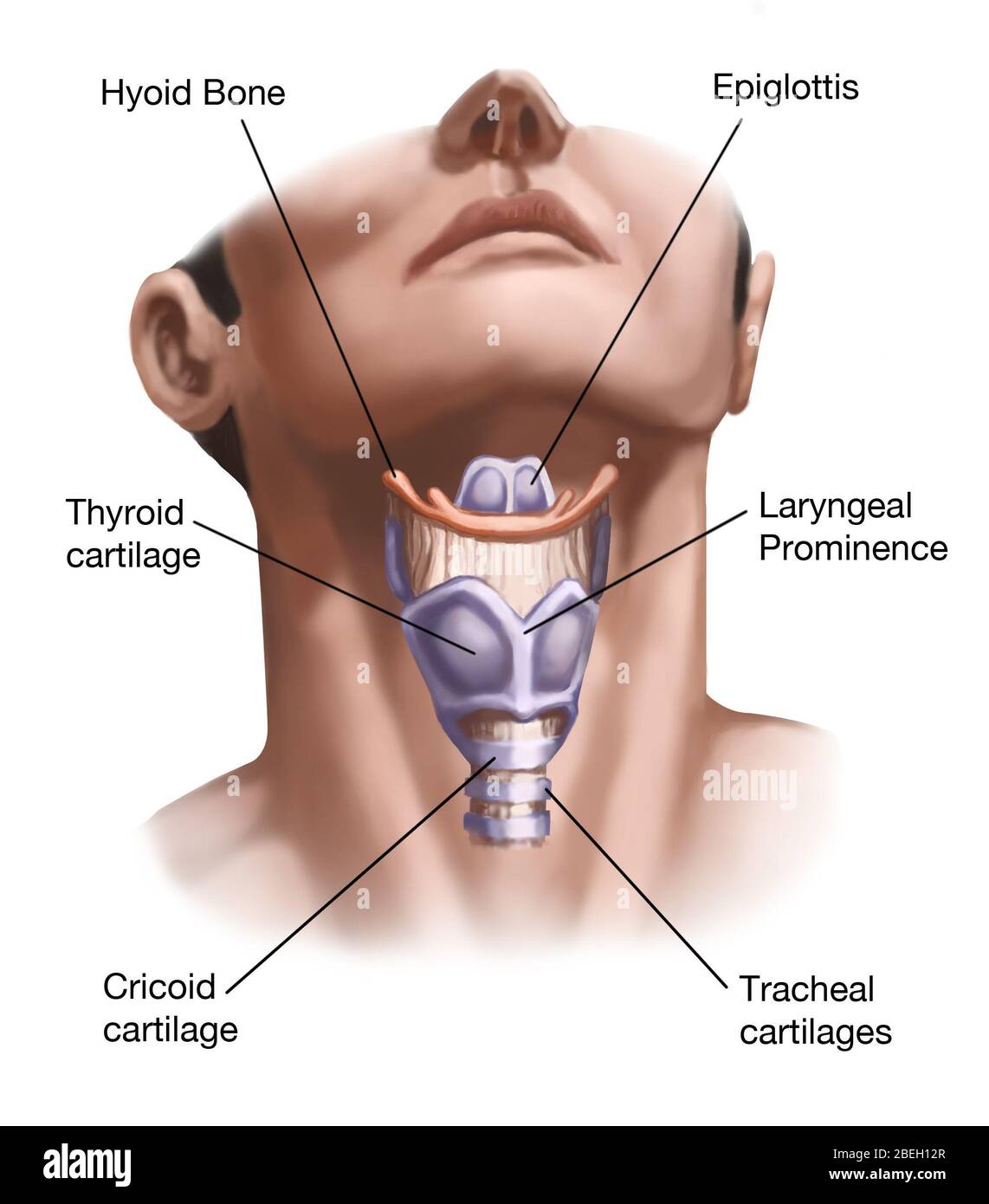
Anatomy Of The Neck Stock Photo Alamy from c8.alamy.com For primary tumors of the nasopharynx, oral cavity and oropharynx, we strongly recommend. Posterior articulation with cricoid cartilage. The superior thyroid vein crosses the artery near its termination, and the middle thyroid vein a little below the level of the cricoid cartilage;
C3/c4 @ superior border of thyroid cartilage.
The thyroid gland is one of the largest and actively functioning endocrine glands. It lies behind the sternohyoid and sternothyroid muscles the thyroid cartilage is composed of two plates called laminae that join in the front at an angle of 90 to 120 degrees. The thyroid cartilage is named for its shape (thyreos (gr): Surgical anatomy a detailed knowledge of thyroid anatomy is a prerequisite for thyroid surgery.
17+ Sacroiliac Joint Ligaments Anatomy . Learn everything about its anatomy and function now at kenhub! The sacroiliac joint (sij) is a synovial joint between ilium and the sacrum. Sacroiliac Joint Pain Pontchartrain Orthodepics Sports Medicine from posm.org Attach the iliac tuberosity and surrounding area to the three sacral fossae. Because the pelvis is a ring, these ligaments work somewhat like the hoops that. Describe the anatomy of the bones, the ligaments, the muscles, and describe the biomechanics of the sacroiliac joint (sij), including coupled movements, normal and abnormal… Our sacroiliac joint exercises will give your pelvis better stability and alleviate your pain. It is the hub for the transference of force between the legs and torso. The posterior sacroiliac ligament is a continuation of interossus sacroiliac ligament. Twisting poses are a top cause of si joint injury. T...
47+ Anatomy Of Calcaneus . Related online courses on physioplus. Want to learn more about it? Anatomy Of The Calcaneus Calcaneus from www.netterimages.com The calcaneus is the largest tarsal bone in the foot and is well designed to sustain high tensile, bending, and compressive forces. The calcaneus is the largest of all the bones in the foot. Want to learn more about it? Formulary drug information for this topic. The calcaneus is the largest of all the bones in the foot. The os calcis is the most frequently. The calcaneus (os calcis) is the largest of the tarsal bones. Calcaneus bone anatomy & function. Source: somepomed.org Assessment of traumatic brain injury online course: Source: lookaside.fbsbx.com Ebraheim's educational animated video describes the anatomy of the calcaneus bone of the foot, and the ...
43+ Surface Anatomy Terminology . Notable anatomic landmarks include the helix, crura, scapha (also known as the scaphoid fossa), antihelix, tragus, antitragus, and lobule (fig. Surface anatomy deals with anatomical features that can be studied by sight, without dissection. Anatomy And Physiology Anatomical Position And Directional Terms from www.visiblebody.com Anatomy is the study of the structure of the body. A delphi consensus convened to create a novel surface anatomy terminology. This unit covers the surface anatomy of the human brain, its internal structure, and the overall organization of sensory and motor systems in the brainstem and spinal cord. Means the caudal(bottom) surface of the manus(front paw) including the carpus (from the antebrachial joint distally). Divided into 9 regions by two vertical and two horizontal imaginary planes divided into 4 quadrants by single verti...


Comments
Post a Comment