32+ Forearm Muscles Anatomy Mri. All superficial muscles are arises from the medial epicondyle of humerus but they are inserted into the different part except. Forearm fractures anatomy and assessment orthopaedicprinciples com.
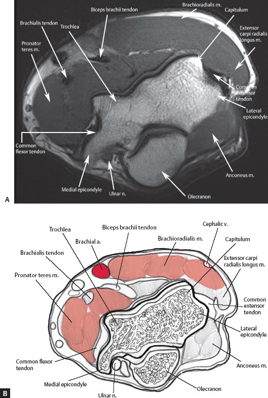
Normal Mri Anatomy Of The Musculoskeletal System Radiology Key from radiologykey.com Musculoskeletal anatomy, kinesiology, and palpation for manual therapists. Myotonic dystrophy, type i arms common: In magnetic resonance imaging (mri) of the elbow, patients are imaged in the supine position or in the prone position with the arm overhead.
Fortunately, there's some patterns that can make the forearm a little bit easier.
3d anatomy tutorial on the muscles of the flexor compartment of the forearm. The brachioradialis is a muscle of the forearm that flexes the forearm at the elbow. Mri of the upper limb. Magnetic resonance imaging (mri) utilizes magnet and radio waves to produce diagnostic images that allow a doctor to visualize the hips.
Comments
Post a Comment