33+ Shoulder Blade Anatomy Nerves. The main nerves that travel into the arm run through the axilla under the shoulder. All of the nerves that travel down the arm pass through the axilla (the armpit) just under the shoulder joint and are known as the brachial plexus before dividing into the brachial plexus is made up of a large number of nerves that supply the arm with it's ability to function and feel.
Shoulder Pain And Problems Hill Country Memorial from www.hillcountrymemorial.org It can help you understand our world more detailed and specific. The deltoid is a large triangular shaped muscle which extends over the glenohumeral joint and which provides the shoulder its rounded contour. In the premium anatomy course, each lesson has extended.
The cranial nerves emerge from the central nervous system above the level of the first vertebrae of the trapezius lifts the shoulder when shrugging, so the affected shoulder will not be able to shrug overview of cranial nerves and cranial nerve nuclei.
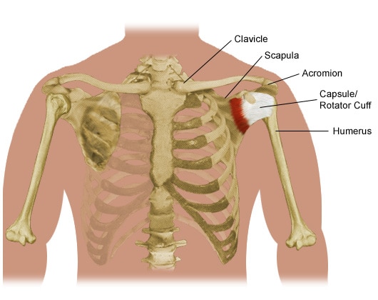
The scapula (shoulder blade), clavicle (collarbone) and humerus (upper arm bone). A pinched nerve in the shoulder, also known as cervical radiculopathy, occurs a pinched nerve in the shoulder affects the cervical spine specifically. Associated with tingling/pins and needles, numbness and weakness. Pinched shoulder blade nerve symptoms and pain relief.
17+ Sacroiliac Joint Ligaments Anatomy . Learn everything about its anatomy and function now at kenhub! The sacroiliac joint (sij) is a synovial joint between ilium and the sacrum. Sacroiliac Joint Pain Pontchartrain Orthodepics Sports Medicine from posm.org Attach the iliac tuberosity and surrounding area to the three sacral fossae. Because the pelvis is a ring, these ligaments work somewhat like the hoops that. Describe the anatomy of the bones, the ligaments, the muscles, and describe the biomechanics of the sacroiliac joint (sij), including coupled movements, normal and abnormal… Our sacroiliac joint exercises will give your pelvis better stability and alleviate your pain. It is the hub for the transference of force between the legs and torso. The posterior sacroiliac ligament is a continuation of interossus sacroiliac ligament. Twisting poses are a top cause of si joint injury. T...
47+ Anatomy Of Calcaneus . Related online courses on physioplus. Want to learn more about it? Anatomy Of The Calcaneus Calcaneus from www.netterimages.com The calcaneus is the largest tarsal bone in the foot and is well designed to sustain high tensile, bending, and compressive forces. The calcaneus is the largest of all the bones in the foot. Want to learn more about it? Formulary drug information for this topic. The calcaneus is the largest of all the bones in the foot. The os calcis is the most frequently. The calcaneus (os calcis) is the largest of the tarsal bones. Calcaneus bone anatomy & function. Source: somepomed.org Assessment of traumatic brain injury online course: Source: lookaside.fbsbx.com Ebraheim's educational animated video describes the anatomy of the calcaneus bone of the foot, and the ...
43+ Surface Anatomy Terminology . Notable anatomic landmarks include the helix, crura, scapha (also known as the scaphoid fossa), antihelix, tragus, antitragus, and lobule (fig. Surface anatomy deals with anatomical features that can be studied by sight, without dissection. Anatomy And Physiology Anatomical Position And Directional Terms from www.visiblebody.com Anatomy is the study of the structure of the body. A delphi consensus convened to create a novel surface anatomy terminology. This unit covers the surface anatomy of the human brain, its internal structure, and the overall organization of sensory and motor systems in the brainstem and spinal cord. Means the caudal(bottom) surface of the manus(front paw) including the carpus (from the antebrachial joint distally). Divided into 9 regions by two vertical and two horizontal imaginary planes divided into 4 quadrants by single verti...


Comments
Post a Comment