20+ Aortic Arch Anatomy Variants. At the top of the ascending aorta, the aorta curves downward in an arch and descends inferiorly (toward the feet) until it reaches the diaphragm, the muscle at the floor of the thorax that separates the thorax from the abdomen. The aortic arch is a continuation of the ascending aorta and begins at the level of the second sternocostal joint.
Neuroscience Files On Twitter Vascularanatomy Endovascularsurgery Stroke Neurosurgery Neurointervention Aortic Arch Anatomy Bovine Variant Common Carotid Origin And Ica Dolichoarteriopathy Tortuosity Kinking Coiling Https T Co from pbs.twimg.com In the mammal the 5th pair do not form. As such, some of the information contained herein. The arch of aorta (aortic arch) is covered anteriorly by the pleura and anterior margins of the lungs, and by the remains of the thymus.
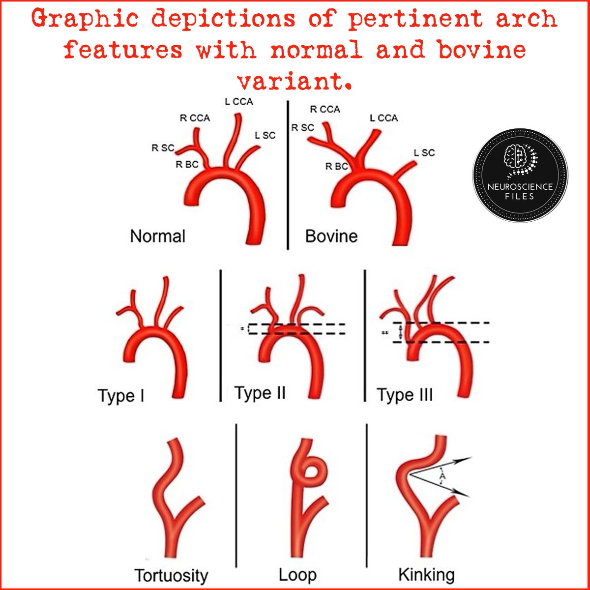
Anatomy of the aortic arches and the great vessels derived therefrom.
Variant anatomy of the aortic arch occurs when there is failure of normal aortic development. Catheter manipulation time (cmt) was assessed for each patient. Normally, the aorta ascends in the superior mediastinum to the level of the sternal notch before arching posteriorly and descending in the left hemithorax. The arch of the aorta or aortic arch (latin:
17+ Sacroiliac Joint Ligaments Anatomy . Learn everything about its anatomy and function now at kenhub! The sacroiliac joint (sij) is a synovial joint between ilium and the sacrum. Sacroiliac Joint Pain Pontchartrain Orthodepics Sports Medicine from posm.org Attach the iliac tuberosity and surrounding area to the three sacral fossae. Because the pelvis is a ring, these ligaments work somewhat like the hoops that. Describe the anatomy of the bones, the ligaments, the muscles, and describe the biomechanics of the sacroiliac joint (sij), including coupled movements, normal and abnormal… Our sacroiliac joint exercises will give your pelvis better stability and alleviate your pain. It is the hub for the transference of force between the legs and torso. The posterior sacroiliac ligament is a continuation of interossus sacroiliac ligament. Twisting poses are a top cause of si joint injury. T...
47+ Anatomy Of Calcaneus . Related online courses on physioplus. Want to learn more about it? Anatomy Of The Calcaneus Calcaneus from www.netterimages.com The calcaneus is the largest tarsal bone in the foot and is well designed to sustain high tensile, bending, and compressive forces. The calcaneus is the largest of all the bones in the foot. Want to learn more about it? Formulary drug information for this topic. The calcaneus is the largest of all the bones in the foot. The os calcis is the most frequently. The calcaneus (os calcis) is the largest of the tarsal bones. Calcaneus bone anatomy & function. Source: somepomed.org Assessment of traumatic brain injury online course: Source: lookaside.fbsbx.com Ebraheim's educational animated video describes the anatomy of the calcaneus bone of the foot, and the ...
43+ Surface Anatomy Terminology . Notable anatomic landmarks include the helix, crura, scapha (also known as the scaphoid fossa), antihelix, tragus, antitragus, and lobule (fig. Surface anatomy deals with anatomical features that can be studied by sight, without dissection. Anatomy And Physiology Anatomical Position And Directional Terms from www.visiblebody.com Anatomy is the study of the structure of the body. A delphi consensus convened to create a novel surface anatomy terminology. This unit covers the surface anatomy of the human brain, its internal structure, and the overall organization of sensory and motor systems in the brainstem and spinal cord. Means the caudal(bottom) surface of the manus(front paw) including the carpus (from the antebrachial joint distally). Divided into 9 regions by two vertical and two horizontal imaginary planes divided into 4 quadrants by single verti...


Comments
Post a Comment