14+ Renal Arteries Anatomy. Renal artery disease renal artery disease is peripheral artery disease that occurs in the blood vessels leading. Each renal artery is about 4 to 6 centimeters long and 5 to 6 millimeters in diameter.
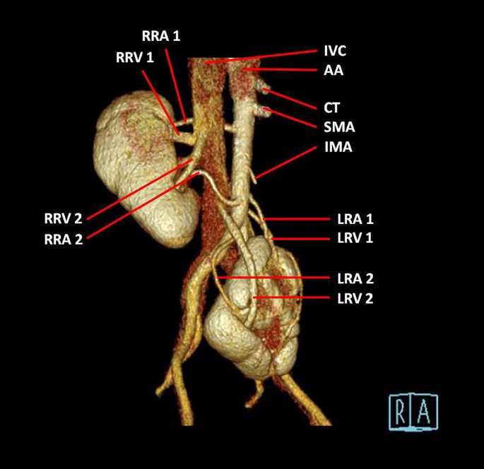
Evaluating The Origin Of Vascular Structures In Ectopic Kidneys With Multidetector Computed Tomography Springerlink from media.springernature.com Arteria renalis) are two lateral branches of the abdominal aorta that arise just inferior to the superior mesenteric artery between vertebrae l1 and l2 and supply the kidneys. The left renal vein separates the left renal artery from the body and tail of the the renal arteries can be involved in many surgical disorders like renal artery aneurysms, renal artery. It is located above the renal vein.
The kidneys are paired organs which reside in the dorsal abdomen.
Renal artery variations including their number, source, and course are very common. The left renal vein separates the left renal artery from the body and tail of the the renal arteries can be involved in many surgical disorders like renal artery aneurysms, renal artery. Supernumerary renal arteries (two or. Stenosis and occlusion are usually due to thromboemboli, atherosclerosis.
17+ Sacroiliac Joint Ligaments Anatomy . Learn everything about its anatomy and function now at kenhub! The sacroiliac joint (sij) is a synovial joint between ilium and the sacrum. Sacroiliac Joint Pain Pontchartrain Orthodepics Sports Medicine from posm.org Attach the iliac tuberosity and surrounding area to the three sacral fossae. Because the pelvis is a ring, these ligaments work somewhat like the hoops that. Describe the anatomy of the bones, the ligaments, the muscles, and describe the biomechanics of the sacroiliac joint (sij), including coupled movements, normal and abnormal… Our sacroiliac joint exercises will give your pelvis better stability and alleviate your pain. It is the hub for the transference of force between the legs and torso. The posterior sacroiliac ligament is a continuation of interossus sacroiliac ligament. Twisting poses are a top cause of si joint injury. T...
27+ Lower Body Anatomy . Free gross anatomy of the human body : Lower body muscle anatomy conclusions. Lower Body Anatomy Artwork Stock Image C014 5581 Science Photo Library from media.sciencephoto.com The anatomical regions of the body. Anatomy of a woman's lower body, anatomy of female lower body, anatomy of lower right side of body, anatomy of the female lower body. Lower body anatomy and color study. Blood vessels and lymphatics of the lower limb. It is composed of many different types of cells that together create tissues and subsequently organ systems. Find the perfect lower body anatomy stock photos and editorial news pictures from getty images. Functional anatomy of the hip. The human body is the structure of a human being. Source: d3uigcfkiiww0g.cloudfront.net Blood vessels and lymphatics of the lower limb. So...
39+ Zebra Anatomy . We hope this picture zebra anatomy in detail can help you study and research. For more anatomy content please follow us and visit our website: What Do Zebra Eat Zebras Diet By Types What Eats Zebras from www.bioexplorer.net Zebra anatomy the zebra has a strong, muscular and symmetrical body, that is zebra anatomy in detail in this image, you will find trachea, esophagus, diaphragm, colon, lungs, heart, stomach, small. Their feet are capped with a singular, hard hoof, which packs a punch when kicked at predators. A glorious fixture of the african landscape, the zebra is a very flashy member of the normally. Zebra anatomy the zebra has a strong, muscular and symmetrical body, that is zebra anatomy in detail in this image, you will find trachea, esophagus, diaphragm, colon, lungs, heart, stomach, small. Although the giraffe looks like an extraordinary animal, it's...
Comments
Post a Comment