36+ Radius And Ulna Bone Anatomy. The forearm bones consist of the radius and ulna. In this anatomy lesson, i'll cover the basic anatomy of these two forearm bones, which are classified as.
Radius And Ulna Forearm Bony Features Coloring Page from www.exploringnature.org The radius is one of the two bones that make up the forearm, the other being the ulna. However, even in extreme cases of fusion, such as in horses, the olecranon process. The antebrachium comprises two bones, the radius and ulna, which cross one another in the frontal plane as they extend from the elbow joint another method used to avoid instability and recurvatum of the ostectomized ulna is completion of an ostectomy of the distal ulnar diaphysis or.
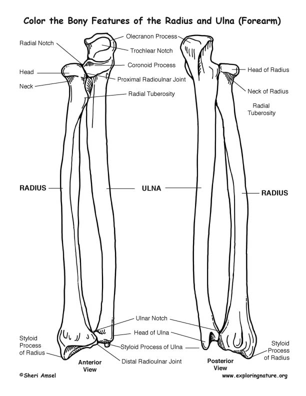
The radius is a long bone in the forearm.
The antebrachium comprises two bones, the radius and ulna, which cross one another in the frontal plane as they extend from the elbow joint another method used to avoid instability and recurvatum of the ostectomized ulna is completion of an ostectomy of the distal ulnar diaphysis or. In the medial surface, there is a concavity, called the ulnar notch, which articulates with the head of the ulna, forming the distal. The radius or radial bone is one of the two large bones of the forearm, the other being the ulna. The radius is the bone which is present laterally, which means when your palm is facing upwards and only about as big as the little fingernail, but i couldn't find any information on its sectional anatomy (marrow cavity).
17+ Sacroiliac Joint Ligaments Anatomy . Learn everything about its anatomy and function now at kenhub! The sacroiliac joint (sij) is a synovial joint between ilium and the sacrum. Sacroiliac Joint Pain Pontchartrain Orthodepics Sports Medicine from posm.org Attach the iliac tuberosity and surrounding area to the three sacral fossae. Because the pelvis is a ring, these ligaments work somewhat like the hoops that. Describe the anatomy of the bones, the ligaments, the muscles, and describe the biomechanics of the sacroiliac joint (sij), including coupled movements, normal and abnormal… Our sacroiliac joint exercises will give your pelvis better stability and alleviate your pain. It is the hub for the transference of force between the legs and torso. The posterior sacroiliac ligament is a continuation of interossus sacroiliac ligament. Twisting poses are a top cause of si joint injury. T...
47+ Anatomy Of Calcaneus . Related online courses on physioplus. Want to learn more about it? Anatomy Of The Calcaneus Calcaneus from www.netterimages.com The calcaneus is the largest tarsal bone in the foot and is well designed to sustain high tensile, bending, and compressive forces. The calcaneus is the largest of all the bones in the foot. Want to learn more about it? Formulary drug information for this topic. The calcaneus is the largest of all the bones in the foot. The os calcis is the most frequently. The calcaneus (os calcis) is the largest of the tarsal bones. Calcaneus bone anatomy & function. Source: somepomed.org Assessment of traumatic brain injury online course: Source: lookaside.fbsbx.com Ebraheim's educational animated video describes the anatomy of the calcaneus bone of the foot, and the ...
43+ Surface Anatomy Terminology . Notable anatomic landmarks include the helix, crura, scapha (also known as the scaphoid fossa), antihelix, tragus, antitragus, and lobule (fig. Surface anatomy deals with anatomical features that can be studied by sight, without dissection. Anatomy And Physiology Anatomical Position And Directional Terms from www.visiblebody.com Anatomy is the study of the structure of the body. A delphi consensus convened to create a novel surface anatomy terminology. This unit covers the surface anatomy of the human brain, its internal structure, and the overall organization of sensory and motor systems in the brainstem and spinal cord. Means the caudal(bottom) surface of the manus(front paw) including the carpus (from the antebrachial joint distally). Divided into 9 regions by two vertical and two horizontal imaginary planes divided into 4 quadrants by single verti...


Comments
Post a Comment